Introduction:
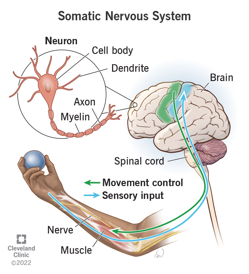
Neurons are the fundamental building blocks of the nervous system, serving as the primary functional units for transmitting and processing information. To accurately understand and study correctly label the following anatomical features of a neuron, it is crucial to correctly identify and label their anatomical features. This article aims to provide a comprehensive guide on correctly labeling the essential components of a neuron.
-
Cell Body (Soma):
The cell body, also known as the soma, is the central region of the neuron. It contains the nucleus, which houses the genetic material of the cell. Correctly labeling the cell body is the first step in identifying a correctly label the following anatomical features of a neuron. Use a clear label to indicate the soma in diagrams or illustrations.
-
Dendrites:
Dendrites are branching extensions that emanate from the cell body. These structures receive signals from other correctly label the following anatomical features of a neuron and transmit them towards the cell body. When labeling dendrites, it is important to differentiate them from axons. Use distinct markings or colors to ensure clarity in diagrams.
-
Axon:
The axon is a long, slender projection that carries nerve impulses away from the cell body toward other correctly label the following anatomical features of a neuron, muscles, or glands. It is crucial to accurately label the axon to distinguish it from dendrites. In illustrations, use a different color or style to highlight the axon.
-
Axon Hillock:
The axon hillock is the region where the axon originates from the cell body. It plays a vital role in the initiation of nerve impulses. Clearly mark the axon hillock when labeling a correctly label the following anatomical features of a neuron to emphasize its significance in the transmission of signals.
-
Myelin Sheath:
The myelin sheath is a fatty, insulating layer that surrounds some axons, facilitating faster transmission of nerve impulses. Label the myelin sheath when it is present, and differentiate it from the axon by using appropriate annotations or shading in diagrams.
-
Nodes of Ranvier:
Nodes of Ranvier are small gaps in the myelin sheath along the axon. They play a crucial role in speeding up the transmission of nerve impulses. When labeling a neuron, mark the Nodes of Ranvier to highlight their functional importance.
-
Synaptic Terminals:
Synaptic terminals, also known as axon terminals, are the end points of the axon where neurotransmitters are released to communicate with other correctly label the following anatomical features of a neuron or target cells. Accurate labeling of synaptic terminals is essential for understanding the communication between neurons.
Conclusion:
Correctly labeling the anatomical features of a anatomical features of a neuron is essential for accurate communication and understanding in the field of neuroscience. Whether creating diagrams for educational purposes or studying neuron structure, attention to detail in labeling the cell body, dendrites, axon, axon hillock, myelin sheath, Nodes of Ranvier, and synaptic terminals is crucial. A clear and accurate representation of these components will contribute to a better comprehension of the intricate functioning of neurons within the nervous system.


